What are the indications for ultrasound during pregnancy?
A prenatal test termed an ultrasound, sometimes known as a sonogram, is provided to most pregnant women. It displays an image of your unborn child within the uterus (womb) using sound waves. Your doctor can monitor the health and growth of your baby with the use of ultrasound.
Pregnancy ultrasounds might be unique because they allow you to "see" your unborn child for the first time. You might be able to see your baby's hands, legs, and other body parts depending on when it's finished and how they're positioned. If you are unsure about the gender of your unborn child, make sure your healthcare practitioner knows! You might be able to tell!
{module id="128"}
Most women get an ultrasound in their second trimester at 18 to 20 weeks of pregnancy. Some also get a first-trimester ultrasound (also called an early ultrasound) before 14 weeks of pregnancy. The number of ultrasounds and timing may be different for women with certain health conditions like as asthma and obesity. Reading about first-trimester ultrasound screenings can help clarify how early scans are used.
Talk to your provider about when an ultrasound is right for you.
What are some reasons for having an ultrasound?
Your provider uses ultrasound to do several things, including:
- To confirm (make sure) you’re pregnant
- To check your baby’s age and growth. This helps your provider figure out your due date.
- To check your baby’s heartbeat, muscle tone, movement and overall development
- To check to see if you’re pregnant with twins, triplets or more (also called multiples)
- To check if your baby is in the heads-first position before birth
- To examine your ovaries and uterus (womb). Ovaries are where eggs are stored in your body.
Ultrasound is another tool that your clinician employs for testing and screening. Screening does not imply conclusively determining whether your child actually has a health condition; rather, it indicates whether your child is more likely than others to have one. Ultrasound may be used by your provider:
- to check for congenital problems such as heart disorders or spina bifida. Following an ultrasound, your doctor can recommend additional testing, known as diagnostic testing, to confirm whether your child has a birth abnormality. A newborn is born with certain health issues, known as birth defects. Birth defects cause one or more body parts to alter in form or function. They may result in issues with general health, the body's development, or its functionality.
- to support additional prenatal testing such as amniocentesis (also known as amnio) or chorionic villus sampling (commonly known as CVS). Placental cells are extracted for testing in CVS. The tissue that supplies your unborn child with nutrition is the placenta. Amniotic fluid and cells are extracted from the sac around your unborn child for the amnio test.
- should look for signs of miscarriage, molar pregnancy, and ectopic pregnancy, among other pregnancy issues.
Are there different kinds of ultrasound?
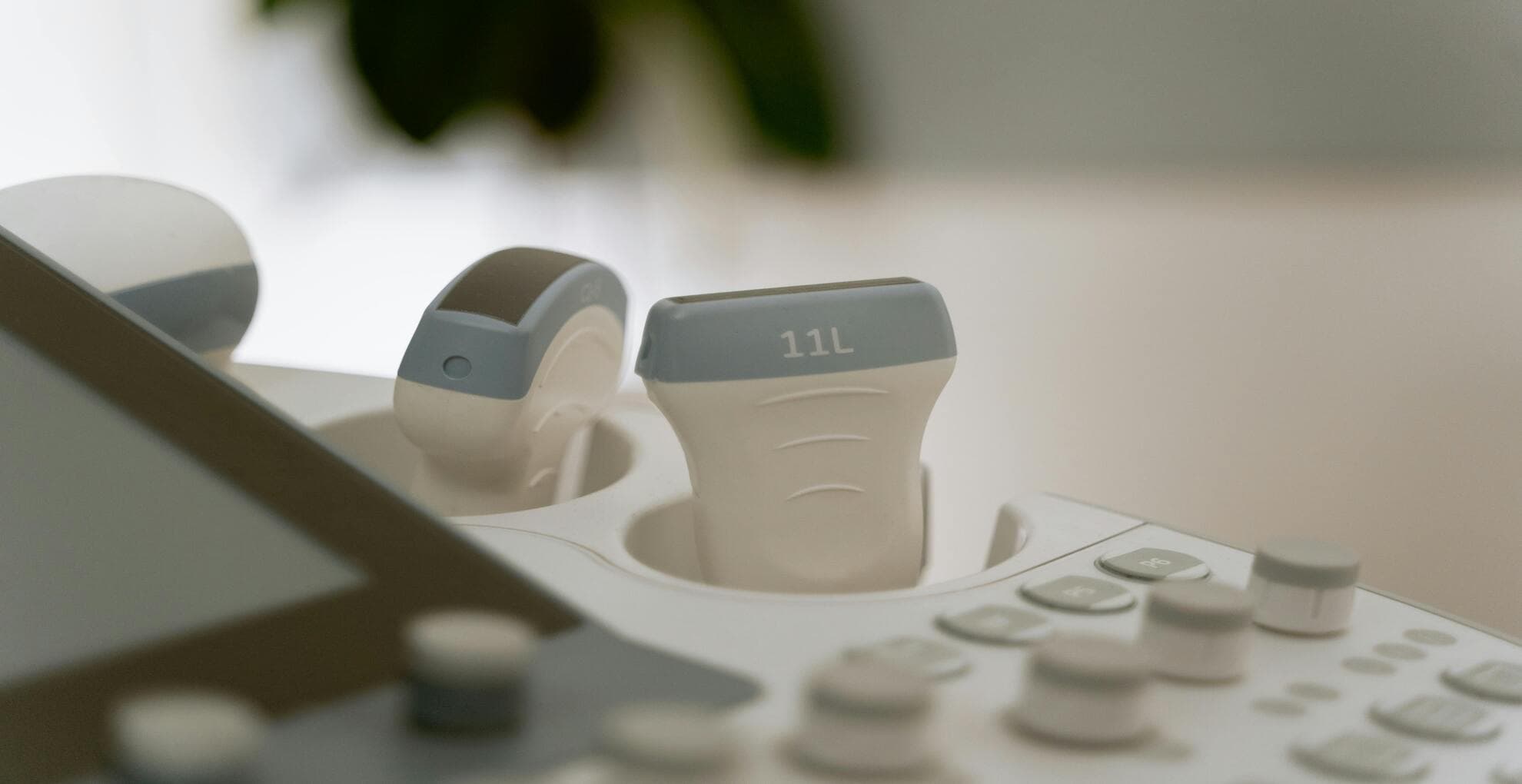
Indeed. The type you receive is determined on the things your doctor is looking for and the stage of your pregnancy. Every ultrasound employs a device known as a transducer, which projects images of your baby onto a computer through the use of sound waves. The most typical ultrasonography types are:
- Transabdominal ultrasound. This type of ultrasound is most likely what you hear about when you are pregnant. Your doctor places a thin coating of gel over your stomach as you lie on your back on an examination table. The gel improves the clarity of the image by facilitating the movement of sound waves. The transducer is then moved around your abdomen by him. In order to have a full bladder during the test, you might need to consume many glasses of water approximately two hours prior to the exam. A full bladder facilitates easier sound wave movement for improved image quality. While having a full bladder can be painful, ultrasounds are painless. A 20-minute ultrasound often takes place.
- Transvaginal ultrasound. The birth canal, or vagina, is used to do this type of ultrasound. You place your feet in stirrups and lie on your back on an examination table. A small, wand-shaped transducer is inserted into your vagina by your healthcare professional. The transducer may exert some pressure on you, but it shouldn't hurt. You should be partially or completely emptied of your bladder. It takes around 20 minutes for this type of ultrasonography as well.
In certain circumstances, your doctor might utilize these types of ultrasound to learn more about your child:
- Doppler ultrasound. If the growth of your baby is abnormal, this type of ultrasound is performed to evaluate the flow of blood to him. In order to assess the blood flow in the umbilical cord and certain blood vessels in your baby as well as to listen to your baby's heartbeat, your provider will use a transducer. If you have Rh illness, a Doppler ultrasound may also be recommended. If left untreated, this blood disorder could have major consequences for your unborn child.
- 3-D ultrasound. A 3-D ultrasound can simultaneously capture thousands of images. It creates an almost photographic-quality 3-D image. In order to ensure that your baby's organs are growing and developing normally, several physicians utilize this type of ultrasound. It can also examine a baby's face for anomalies. A three-dimensional ultrasound could also be performed to look for uterine issues.
- 4-D ultrasound. Similar to a three-dimensional ultrasound, this allows you to see your baby's movements on video.
Does ultrasound have any risks?
When your healthcare professional performs ultrasounds, you and your unborn child are protected. Ultrasound is safer than X-rays since it employs sound waves rather than radiation. After using ultrasonography for more than 30 years, providers have not discovered any serious hazards.
Ultrasound is useful in ruling out issues if your pregnancy is healthy, but it is not a perfect tool. It might not catch all birth defects. Occasionally, a regular ultrasound may reveal a birth defect when none actually exists. Even while follow-up exams frequently reveal the baby's health, parents may become concerned when there are false alarms.
You might be aware of certain establishments that provide parents with “keepsake” 3-D or 4-D ultrasound images or movies, such as shops in a mall, but aren't owned by physicians or other health care providers. These non-medical ultrasounds are not advised by the Food and Drug Administration (FDA), the American College of Obstetricians and Gynecologists (ACOG), or the American Institute of Ultrasound in Medicine (AIUM). They might not be medically trained, and the information they give you could be harmful or inaccurate.
What are the benefits of ultrasound during pregnancy?
Ultrasonography typically reveals that the baby is growing normally in most women. As long as your ultrasound results are normal, make sure to continue with your prenatal visits. If you'd like a clearer picture of how growth is measured, reviewing fetal size by week can offer helpful context.
An ultrasound may occasionally reveal that you and your child require particular attention. Your kid might receive treatment in the womb before to delivery, for instance, if the ultrasound reveals that he has spina bifida. Your healthcare practitioner may attempt to turn your baby's position to head-down if the ultrasound indicates that they are breech, or they may need to perform a cesarean section, often known as a c-section.
{module id="129"}
An ultrasound may occasionally reveal that you and your child require particular attention. Your kid might receive treatment in the womb before to delivery, for instance, if the ultrasound reveals that he has spina bifida. Your healthcare practitioner may attempt to turn your baby's position to head-down if the ultrasound indicates that they are breech, or they may need to perform a cesarean section, often known as a c-section.
If you are worried about going to the doctor related to your pregnancy, try Soula. Soul is AI assistant for Female Wellbeing. Soula has been specifically designed for women, taking their unique challenges on their journey towards success and joy. Soula is here 24/7 to provide women with emotional and informational support during the most challenging periods of their lives, aiming to prevent anxiety, burnout, and depression through neuroscience techniques and conversational AI.
How many times should I have an ultrasound during pregnancy?
Two ultrasounds are usually necessary for a healthy pregnancy: one at about 11 to 14 weeks and another at about 18 to 20 weeks. Many parents also read about second-trimester screening to understand how the mid-pregnancy scan fits into overall prenatal care.
You may require additional testing if any abnormalities or problems are found during either of the standard ultrasounds.
During the last month of pregnancy, weekly ultrasounds may be advised in high-risk pregnancies, which are typically caused by the weight, age, or medical history of the mother. If your provider suggests additional scans later in pregnancy, the overview of third-trimester screening may help you understand what to expect.